Posts

Showing posts from December, 2022

Study of compound microscope

Image
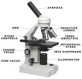
  AIM : Study of compound microscope INTRODUCTION : A compound microscope is a type of microscope that uses two sets of lenses to magnify the image under the microscope. it has an objective lens that has a resolution of 4x,10x, 40x, 100x, and an eyepiece of resolution of 10x. the usage of both lenses, compounds or magnifies the image of the object below the objective lens to achieve a resolution of 40x, 100x, 400x, 1000x. Parts of the compound microscope may be grouped into two major categories   Ø   Mechanical parts Ø   Optical parts.   (a)   Mechanical Parts:   These include base or foot, pillar, arm, inclination joint, stage, clips, diaphragm, body tube, nose piece, coarse adjustment knob and fine adjustment knob. (b)   Optical Parts:   These include eye piece lens, objective lenses and mirror. All these parts are briefly described below: 1.   Foot or Base: It is the basal, horse shoe-

Electrophoresis Principle and its types

Image
  Electrophoresis   Electrophoresis is a technique used to separate macromolecules in a fluid or gel based on their charge, binding affinity, and size under an electric field. In the year 1807, Ferdinand Frederic Reuss was the first person to observe electrophoresis. He was from Moscow State University. Anaphoresis is the electrophoresis of negative charge particles or anions whereas cataphoresis is electrophoresis of positive charge ions or cations.  Electrophoresis  has a wide application in separating and analysing biomolecules such as proteins, plasmids, RNA, DNA, nucleic acids. Electrophoresis Principle and its types: Charged macromolecules are placed in the electric field move towards the negative or positive pole based on their charge. Nucleic acid has a negative charge and therefore it migrates towards the anode. This technique is divided into two types viz slab electrophoresis and capillary electrophoresis. Types of Electrophoresis: 1.        Capillary electrophor

ਪੀਲੀਆ

ਪੀਲੀਆ ਪੀਲੀਆ ਇੱਕ ਬਿਮਾਰੀ ਹੈ ਜੋ ਚਮੜੀ, ਸਕਲੇਰੀ (ਅੱਖ ਦਾ ਚਿੱਟਾ ਹਿੱਸਾ) ਅਤੇ ਹੋਰ ਲੇਸਦਾਰ ਝਿੱਲੀ ਦੇ ਪੀਲੇ ਰੰਗ ਦਾ ਕਾਰਨ ਬਣਦੀ ਹੈ। ਇਸ ਤੋਂ ਇਲਾਵਾ, ਸਰੀਰ ਦੇ ਤਰਲ ਵੀ ਪੀਲੇ ਰੰਗ ਵਿੱਚ ਬਦਲ ਸਕਦੇ ਹਨ। ਤਕਨੀਕੀ ਤੌਰ 'ਤੇ, ਇਸ ਨੂੰ icterus ਵੀ ਕਿਹਾ ਜਾਂਦਾ ਹੈ। ਇਹ ਖੂਨ ਅਤੇ ਸਰੀਰ ਦੇ ਟਿਸ਼ੂਆਂ ਵਿੱਚ ਬਿਲੀਰੂਬਿਨ ਦੇ ਇਕੱਠੇ ਹੋਣ ਕਾਰਨ ਹੁੰਦਾ ਹੈ। ਬਿਲੀਰੂਬਿਨ ਇੱਕ ਰਹਿੰਦ-ਖੂੰਹਦ ਉਤਪਾਦ ਹੈ ਜੋ ਉਦੋਂ ਪੈਦਾ ਹੁੰਦਾ ਹੈ ਜਦੋਂ ਲਾਲ ਖੂਨ ਦੇ ਸੈੱਲ ਟੁੱਟ ਜਾਂਦੇ ਹਨ। ਫਿਰ ਇਸਨੂੰ ਖੂਨ ਦੇ ਪ੍ਰਵਾਹ ਰਾਹੀਂ ਜਿਗਰ ਤੱਕ ਪਹੁੰਚਾਇਆ ਜਾਂਦਾ ਹੈ ਜਿੱਥੇ ਇਸਨੂੰ ਫਿਰ ਇੱਕ ਪਾਚਨ ਤਰਲ ਨਾਲ ਮਿਲਾਇਆ ਜਾਂਦਾ ਹੈ ਜਿਸਨੂੰ ਬਾਇਲ ਕਿਹਾ ਜਾਂਦਾ ਹੈ। ਆਮ ਤੌਰ 'ਤੇ, ਬਿਲੀਰੂਬਿਨ ਸਟੂਲ ਦੁਆਰਾ ਡਿਸਚਾਰਜ ਕੀਤਾ ਜਾਂਦਾ ਹੈ ਅਤੇ ਬਾਕੀ ਬਚਿਆ ਪਿਸ਼ਾਬ ਰਾਹੀਂ ਬਾਹਰ ਨਿਕਲਦਾ ਹੈ। ਪਰ ਜੇਕਰ ਬਿਲੀਰੂਬਿਨ ਨੂੰ ਜਿਗਰ ਰਾਹੀਂ ਪ੍ਰਸਾਰਿਤ ਨਹੀਂ ਕੀਤਾ ਜਾ ਸਕਦਾ, ਤਾਂ ਇਹ ਖੂਨ ਵਿੱਚ ਜਮ੍ਹਾ ਹੋ ਜਾਂਦਾ ਹੈ ਜਿਸ ਨਾਲ ਪੀਲੀਆ ਹੋ ਜਾਂਦਾ ਹੈ। ਪੀਲੀਆ ਦੇ ਕਾਰਨ ਅਤੇ ਲੱਛਣ ਪੀਲੀਆ ਨਵਜੰਮੇ ਬੱਚਿਆਂ ਵਿੱਚ ਵਧੇਰੇ ਆਮ ਹੁੰਦਾ ਹੈ ਅਤੇ ਇਸਨੂੰ ਨਵਜੰਮੇ ਪੀਲੀਆ ਕਿਹਾ ਜਾਂਦਾ ਹੈ। ਇਹ ਮੁੱਖ ਤੌਰ 'ਤੇ ਇਸ ਲਈ ਹੁੰਦਾ ਹੈ ਕਿਉਂਕਿ ਇੱਕ ਨਵਜੰਮੇ ਬੱਚੇ ਦਾ ਜਿਗਰ ਪੂਰੀ ਤਰ੍ਹਾਂ ਵਿਕਸਤ ਨਹੀਂ ਹੁੰਦਾ ਹੈ ਅਤੇ ਇਹ ਖੂਨ ਤੋਂ ਬਿਲੀਰੂਬਿਨ ਨੂੰ ਪ੍ਰੋਸੈਸ ਕਰਨ ਵਿੱਚ ਘੱਟ ਪ੍ਰਭਾਵ